Researchers at MIT Lincoln Laboratory and their collaborators at the Center for Ultrasound Research and Translation (CURT) at Massachusetts General Hospital (MGH) have developed a new medical imaging device: non-contact laser ultrasound (NCLUS). This laser-based ultrasound system provides images of internal features such as organs, fat, muscles, tendons and blood vessels. The system can also measure bone strength and potentially track disease stages over time.
“Our patented skin-safe laser system concept aims to transform medical ultrasound by overcoming the limitations associated with traditional contact probes,” explained principal investigator Robert Haupt, a senior researcher in Lincoln Laboratory’s Active Optical Systems Group. Haupt and senior staff member Charles Wynn are co-inventors of the technology, and assistant group leader Matthew Stowe provides technical leadership and oversight of the NCLUS project. Rajan Gurjar is the systems integrator principal, working with Jamie Shaw, Bert Green, Brian Boitnott (now at Stanford University), and Jake Jacobsen on optical and mechanical engineering and system building.
Medical Ultrasound Practice
If your doctor orders an ultrasound, you can expect a trained sonographer to press and operate a set of transducers set up in a handheld device onto your body. When the sonographer pushes the transducer probe through your skin, high-frequency sound waves (ultrasound) penetrate and travel to your body tissues, where they “reverberate” to different tissue structures and characteristics. These echoes appear as changes in acoustic impedance or tissue strength (tissue softness or stiffness) and originate from fat, muscles, organs, blood vessels and bones deep within the body. The probe receives the returning echoes, which are combined into a representative image of the internal features of the body. Specialized processing schemes (synthetic aperture processing) are used to construct the shapes of 2D or 3D tissue features, which are then displayed on a computer monitor in real time.
Using ultrasound, doctors can non-invasively “see” inside the body, imaging different tissues and their geometry. Ultrasound can also measure pulses of blood flow through arteries and veins and can characterize the mechanical properties of tissues and organs (elastography). Ultrasound is commonly used to assist doctors in assessing and diagnosing a variety of health conditions, diseases, and injuries. For example, ultrasound can be used to image the anatomy of a developing fetus, detect tumors, and measure the extent of heart valve stenosis or leakage. From handheld devices on iPhones to cart-based systems, ultrasound is highly portable, relatively inexpensive, and widely used in both point-of-care and remote field settings.
Limitations of Ultrasound
Although state-of-the-art medical ultrasound systems can resolve tissue features within fractions of a millimeter, the technology has some limitations. Using the sonographer’s bare hands to manipulate the transducer to obtain an optimal viewing window into the body can lead to imaging errors. More specifically, when sonographers apply pressure to a probe by feeling, they randomly compress the local tissue that the probe contacts, causing unpredictable changes in tissue properties that affect the propagation path of ultrasound waves. This compression distorts the image of tissue features and creates a degree of unpredictability, meaning feature shapes cannot be accurately mapped. Additionally, tilting the probe, even slightly, changes the angular plane of the image view—tilting the image and creating uncertainty in the localization of body features.
Image distortion and positional reference uncertainty are large enough that ultrasound cannot resolve with sufficient confidence, for example, whether the tumor is getting larger or smaller and the precise location of the tumor within the host tissue. Furthermore, uncertainties in feature size, shape, and location can change with repeated measurements, even when the same sonographer attempts to retrace their steps. This uncertainty (called operator variability) is exacerbated when different sonographers attempt the same measurement, resulting in interoperator variability. Because of these shortcomings, ultrasound often cannot track cancerous tumors and other disease states. In contrast, methods such as magnetic resonance imaging (MRI) and computed tomography (CT) must track disease progression, even though they are much more costly, have larger system size and complexity, and carry radiation risks.
“Variability has been a major limitation of medical ultrasound for decades,” said Anthony Samir, associate chief of imaging sciences in the MGH Department of Radiology and director of CURT. Samir and his MGH CURT colleagues Kai Thomenius and Marko Jakolvejic provide important medical experience, technology expertise, providing guidance to laboratory teams on conventional ultrasound equipment and working with them to develop the NCLUS system.
By fully automating the process of acquiring ultrasound images, NCLUS has the potential to reduce the need for sonographers and reduce operator variability. Laser positioning allows for accurate reproduction, eliminating variations from repeated measurements. Since the measurement is non-contact, no local tissue compaction or its associated distortion of image features occurs. Additionally, similar to MRI and CT, NCLUS provides a fixed reference frame feature that uses skin markers to reproduce and compare repeated scans over time. To support this tracking capability, the lab team developed software that processes ultrasound images and detects any changes between them. NCLUS requires neither manual pressure nor coupling gel (as required with contact probes) and is also ideal for patients with painful or sensitive areas of the body, in fragile conditions, or at risk for infection.
“NCLUS can image burn or trauma victims, patients with direct access to deep tissue areas during surgery, premature infants requiring intensive care, patients with neck and spine injuries, and distant infectious individuals,” Haupt said.
photoultrasound
NCLUS uses a pulsed laser to transmit light energy to the skin surface through the air. Once the light enters the skin, it is quickly absorbed. The light pulse causes instantaneous local heating and rapidly deforms the skin through a thermoelastic process, which in turn generates ultrasound and acts as an ultrasound source, a phenomenon known as photoacoustics.
The light pulses generate sufficient ultrasound power at a frequency comparable to that of medical ultrasound in practice, while causing no sensation on the skin. The team patented the selection of optical carrier wavelengths and their photoacoustic process is designed to create a consistent source of ultrasound that is not affected by skin color or tissue roughness.
Ultrasound echoes returning from within the tissue appear as localized vibrations on the skin surface, which are measured by a highly sensitive, specialized laser Doppler vibrometer.
“With appropriate laser emission and reception implementations, any exposed tissue surface can become a viable ultrasound source and detector,” Haupt explains.
Advances in clinical operating systems
In 2019, the team demonstrated that the NCLUS proof-of-concept (GEN-1) system could collect ultrasound images from the human body using a skin-safe laser, a first in the medical community. However, acquisition of image data from patient subjects is time-consuming and impractical for clinical practice. Furthermore, the image resolution of the GEN-1 system is significantly lower than that of state-of-the-art medical ultrasound images.
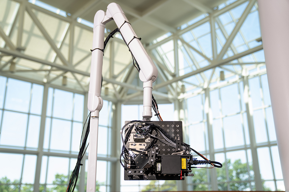
Significant engineering development has since occurred to transform NCLUS GEN-1 into an operating system suitable for clinical testing. In the clinical NCLUS system, both the laser source and receiver are miniaturized and mounted within an optical head attached to a portable armature. The pulsed and scanned laser is 500 times faster than the GEN-1 system, reducing overall image data acquisition time to less than a minute. Future NCLUS prototypes will involve faster acquisition times of less than one second. The new clinical system can also operate at much higher ultrasound frequencies than the GEN-1 system, with resolution as low as 200 microns, which is comparable to the resolution of state-of-the-art medical ultrasound.
The movable armature allows multiple degrees of freedom to view various areas of the body. There are also programmable fast-turning mirrors inside the optical head that automatically position the source and receive the laser beam to accurately build the ultrasonic array. 2D lidar is used to map the patient’s skin surface topography; a high frame rate shortwave infrared camera records the projected position of the laser source and receiver on the skin, providing the array parameters needed to construct an ultrasound image. Skin surface topography mapping and laser position recording are recorded using natural skin features such as freckles. In this way, a fixed frame of reference is established for performing precise repeat scans over time.
The NCLUS Clinical System generates fully automated and registered ultrasound images through synthetic aperture processing. The team demonstrated the system on a gel-based ice sphere that was synthesized to match the mechanical properties of human tissue (called a phantom) that control the propagation of ultrasound waves.
Through sponsored projects, the team is currently developing NCLUS to support cutting-edge military applications. These applications include detecting and characterizing life-threatening injuries caused by intra-organ bleeding; monitoring debilitating musculoskeletal injuries and their healing over time; providing elastography of soft tissue and bone in amputee limb regions to accelerate prosthetic socket development Design and installation. Civilian applications include imaging in intensive care units. With NCLUS, emergency medical technicians, paramedics, and medical personnel without specialized sonography training may be able to perform ultrasound imaging outside the hospital setting—in a doctor’s office, at home, or in a remote battlefield environment.
“With further development, NCLUS has the potential to become a transformative technology: an automated, portable ultrasound platform with fixed reference frame capabilities similar to MRI and CT,” said Samir.
In the next phase of the NCLUS project, the team will conduct clinical studies using an operable, skin-safe laser to evaluate and compare ultrasound images with traditional medical ultrasound images. If these studies are successful, the team will seek commercial funding for clinical medical device development, followed by approval from the U.S. Food and Drug Administration agency.
This work was funded by the U.S. Army Military Combat Medicine Research Program. In vivo testing in humans was approved by the MIT Committee on Human Subjects.