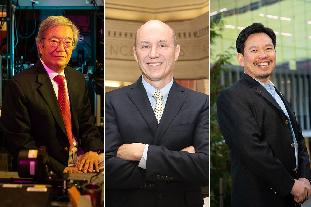
The Lasker Foundation honors James Fujimoto ’79, SM ’81, PhD ’84, Elihu Thomson Professor of Electrical Engineering and Principal Investigator of the Research Laboratory of Electronics (RLE) as the 2023 Lasker-DeBakey Clinical Medical Research Award recipient Pioneering work in optical coherence tomography. Fujimoto collaborates with Eric Swanson SM ’84, a research affiliate of the MIT Research Laboratory of Electronics and a mentor at MIT’s Deshpande Center for Technology Innovation, and David Huang ’85, SM ’89, PhD ’93, professor of ophthalmology at Oregon State Share this award with the University of Health and Sciences.
Considered one of the most prestigious awards in biomedical research, the Lasker Award honors individuals who have made “significant advances in the understanding, diagnosis, treatment, cure, and prevention of human disease.” A large number of Lasker Prize winners went on to win Nobel Prizes.
According to the Lasker Foundation recognition, Fujimoto, Huang and Swanson “are honored for the invention of optical coherence tomography (OCT), a technology that revolutionized the field of ophthalmology and allowed rapid detection of vision-impairing retinal diseases.” Available here An animated video describing the work.
“I am honored to be one of the recipients of this award,” Fujimoto said. “OCT represents a decades-long multidisciplinary collaborative effort by scientists, engineers, the clinical community and industry. We are excited for the opportunity to help improve patient care and extend our sincere thanks to the Lasker Foundation.”
Before the invention of OCT, standard methods for diagnosing eye disease were limited. In the early 1990s, Fujimoto, an electrical engineer and expert in advanced laser technology, teamed up with satellite communications engineer Swanson (then at MIT Lincoln Laboratory) and M.D. student Huang to devise a better way to diagnose the disease. Using an optical technique called interferometry, they have developed for the first time a technique that can image the three-dimensional microstructure of the living retina.
Their work was published in 1991 science, revolutionized the field of ophthalmology and provided more precise methods to detect disease and monitor treatment. Other co-authors of this article include Charles P. Lin, Joel S. Schuman, William G. Stinson, Warren Chang, Michael R. Hee, Thomas Flotte, Kenton Gregory, and Carmen A. Puliafito.
Revolutionizing ophthalmology with the echo of light
To understand how optical coherence tomography works, it is useful to consider other imaging methods that use echoes. “OCT is an optical analog of ultrasound or radar,” explains Fujimoto. “Instead of measuring sound, it measures the echo delay of reflected or scattered light to allow in situ imaging of subsurface microstructures in tissue or materials.”
The short wavelength of light allows OCT to produce images with microscopic resolution, but using light, as opposed to sound, which travels slower and has longer wavelengths, poses thorny technical problems.
“The speed of light is very fast,” Fujimoto points out. “Light from the Moon travels to Earth in 1.3 seconds. So to measure echo time delays at very small scales in biological tissue, you need extremely high-resolution measurement techniques.”
Here, Fujimoto, Swanson and Huang found that their diverse backgrounds enhanced their problem-solving abilities.
“OCT adopts many advanced technologies in the field of high-speed optical communications,” explains Fujimoto. One of the team’s realizations was that infrared light penetrates human tissue well, and interferometry could achieve the required high resolution and sensitivity. This makes it possible to measure the “echo time” of reflected or scattered infrared light waves, creating microscopically high-resolution three-dimensional images of subsurface structures within tissue.
Perform optical biopsy
Importantly, this technology is not a replacement for ultrasound, CT, or MRI, but rather a different tool with unique and complementary advantages. MRI, CT, and ultrasound can penetrate deeply into the body to create full-body images, but the resolution is limited. OCT can perform “optical biopsies,” imaging subsurface structures at microscopic resolution without the need to remove and process specimens. OCT has limited imaging depth in tissues outside the eye, but can be combined with other optical instruments to image within the body.
OCT would not have been developed without interdisciplinary collaboration with clinical scientists. Carmen Puliafito and Joel Schuman, then at the New England Eye Center and Tufts University School of Medicine respectively, led the first effort to develop OCT to treat diabetic retinopathy, age Clinical studies of related macular degeneration and glaucoma. These studies can help determine future clinical applications and commercialization of OCT in ophthalmology.
Retinal imaging became the largest application of OCT; in ophthalmologist offices around the world, it is now considered the standard of care for diagnosing and monitoring eye disease. OCT can also help improve understanding of disease mechanisms and accelerate the development of new drug treatments.
Many ophthalmologists say OCT allows laypeople to detect disease with a sensitivity approaching that of professionals. Conditions such as diabetic retinopathy, age-related macular degeneration and glaucoma may not produce noticeable symptoms in their early stages but can be detected and treated before irreversible vision loss occurs.
Applications of OCT are now being developed for use by the wider public beyond eye clinics. “In the future, diseases can be screened through automated OCT exams at your local pharmacy. The eyes are the window to health – in addition to eye diseases that impair vision, OCT can detect systemic diseases such as diabetes and neurological disorders. For Public Health The impact could be huge,” Fujimoto explained.
OCT also has applications well beyond ophthalmology. The team soon realized that fiber optics could be used to extend OCT coverage to deeper areas of the body, imaging through catheters, endoscopes and laparoscopes.
Intravascular imaging, the second largest application of OCT, was developed in collaboration with cardiologist Mark Brezinski of Massachusetts General Hospital and Harvard Medical School. Brezinski demonstrated that OCT could detect unstable atherosclerotic plaques that lead to heart attacks and led many of the studies that first demonstrated OCT for optical biopsies.
“There are tissues that typically do not require biopsy, such as the retina, coronary arteries, nerves and brain, where OCT can provide in situ pathology information in real time,” Fujimoto said. “Another application is surgical guidance – you can see underneath the tissue surface before making an incision to avoid sensitive nerves and blood vessels.”
Many research groups and clinics are developing technologies and applications, and OCT serves as a shining example of the potential for interdisciplinary and international scientific collaboration. “Interdisciplinary collaboration is very popular now, but was relatively uncommon in the 1990s when OCT was first developed,” explains Fujimoto.
For Fujimoto, OCT’s success and its growing range of applications are a powerful reminder of the importance of interdisciplinary work. “In medicine, as well as many other fields, there is an increasing use of technology, including advanced hardware and analytics and artificial intelligence. Modern medicine can leverage these technologies to improve patient care and reduce mortality.”